Image Quality Described as Close or Equivalent to CT-SIM Scans
As treatment protocols become more demanding, radiation therapy teams require higher image quality to support their treatment decisions. Varian’s technology for iterative cone-beam CT (iCBCT) at the treatment machine meets this need by improving on traditional image reconstruction methods. With iCBCT, advanced algorithms reduce noise and artifacts, yielding higher quality images than was possible with earlier standard approaches to CBCT.
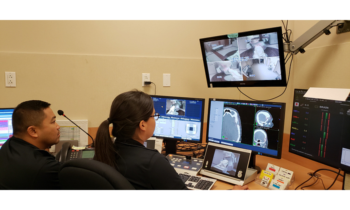
Jon Lagua and Tanya Chavez, radiation therapists, review images at St. Joseph Hospital.
“The quality compared to a diagnostic CT scan is very similar,” said Rob Ash, medical director/radiation oncologist at St. Joseph Hospital in Orange, California. His colleague, chief medical physicist James (Jim) Du, agreed. “iCBCT definitely comes a lot closer to CT-simulation (CT-SIM) quality,” he said, adding that better imaging can facilitate tighter image matching and more accurate patient and target positioning.
iCBCT, currently a standard offering on Varian’s Halcyon™ and Ethos™ platforms and an option on the TrueBeam® and EdgeTM systems, employs two innovative algorithms —1) the Acuros® CTS advanced dose calculation to remove scatter artifacts, and 2) an advanced statistical reconstruction algorithm—plus a high-speed graphics processing unit (GPU)—to quickly generate high-quality images with improved Hounsfield unit uniformity compared to standard CBCT reconstruction methods.
According to Eric Simmons, clinical marketing manager at Varian, iCBCT was developed with the goal of improving the image quality achieved with standard CBCT. Currently, CBCT is primarily used for bone and soft tissue matching during online setup correction at the start of a radiotherapy session. However, streaks, artifacts, scatter, and noise typically seen in standard CBCT scans can make them less useful. iCBCT images offer the sharpness and distinct soft tissue boundaries needed for improved image interpretation.
“The greys for the bladder or prostate, and seminal vesicles are a lot more distinct from the blacks. They pop out more, and the whites are a lot sharper,” noted Hoang Tran, radiation therapist at St. Joseph Hospital. Currently, iCBCT is primarily used for scans of stationary anatomy such as the head, neck, and pelvis.
iCBCT allows better identification of anatomical structures that can be difficult or impossible to identify with a standard CBCT due to scan artifacts. These artifacts can have many causes, from the presence of prostheses to tumor deformation from digestive gas.
“When we scan a patient with an artificial hip or someone who has gas in the pelvic area, we see tremendous improvement in the visualization of anatomy with iCBCT compared to standard CBCT. You have to see it to appreciate it,” remarked Du.
John Seward, radiation therapist at St. Joseph Hospital, described a case where iCBCT was particularly helpful for working with a patient with prostheses.
“We had a patient with bilateral hip implants who was receiving treatment to his prostate and seminal vesicles,” said Seward. “We did an iCBCT scan and the images actually looked better than our CT-SIM scan because the artifacts were removed. We were able to visualize the prostate better than was possible during the planning process. We actually took the iCBCT image and registered it to our planning CT to confirm that the dose was being delivered to the correct location.”
The clinical team at St. Joseph Hospital agrees that the ability to match soft tissue or tumor volume instead of relying on bony anatomy surrogates has the potential to improve the precision of patient setup and potentially spare organs at risk (OAR).
“When we’re doing our treatment planning, the target, which is generally in soft tissue, often moves with respect to the bone, so you can’t get accurate setups by just matching the bones,” remarked Kali Mather, medical physicist at St. Joseph Hospital. “You need to be able to visualize and match soft tissue, so you know you’re hitting the target.”
“iCBCT helps us distinguish between the small bowel and other critical structures, allowing us to spare normal tissues in our postoperative gynecologic patients. It’s helpful to clearly delineate the patient’s anatomy in order to reduce radiation-induced toxicity and side effects,” noted Tanuja Bhandari, radiation oncologist.
Du added, “Better visualization with iCBCT gives us more confidence we’re hitting the target. This is especially helpful in small margin cases, such as SRS, where reducing the setup variability is critical.”
Varian’s iCBCT solution improves image reconstruction efficiency, making it possible to use the technology in an online clinical setting without inconvenient tradeoffs. The advanced algorithms and the high performance, state-of-the-art GPU ensure that iCBCT adds only 10 to 30 seconds to the process.
“The time needed to acquire an iCBCT image is pretty minimal, compared to a CBCT scan,” said Seward. “It’s an additional 20 seconds for the reconstruction.” Though additional time is needed, the improved soft tissue visualization can result in reductions in time spent on patient positioning, image matching, and the need to run multiple scans. The result is often an overall time savings.
“With CBCT scans, it not only takes longer to set the patient up, but you sometimes have to take multiple images,” said Du. “With iCBCT, patient discomfort, time for setup, and CBCT dose is reduced because, most of the time, only one iCBCT image is needed. We definitely see a reduction in the imaging and setup time with iCBCT.”
“If, for example, anatomy is obscured by a gas bubble, we’d have to take the patient down and put them back on the table after the issue is resolved,” added Mather. “So, iCBCT saves both dose and time. It’s also really nice that the image acquisition process with iCBCT is the same as regular CBCT, so you don’t have to put the patient through anything different than the standard care.”
The information captured herein represents the genuine experience of the attributed individuals and may not necessarily represent the views of Varian or the above referenced institutions. Your results may vary. Individuals were not compensated for their participation.